domingo, 11 de septiembre de 2011

The Spleen and Ovulation


At the start of my college education while taking Biology, I was briefly introduced into a very light "view" of what goes on in a woman's body during the ovulation and menstruation period and it may seem corny, but the ability to give life that women posses is just incredible right from the start and lately, I have been reminded of this awesome capacity by some recent births (wich hands down, I'm sure will be some kick ass babies in no time!). A couple of years ago during our Physiology courses, we entered the world of Endocrinology wich included the Female Sex Hormones and that became a pretty exciting topic for me since this time around I had the chance to get a more in depth view on this subject, however the same way that it happens with a bunch of stuff in the Medicine world, a lot of things still go unanswered when it comes to the ovulation phase.

So a couple of days ago, I was "wasting time" jumping through some medical news headlines and I ended up in an Endocrinology journal since we are taking that subjec this semester ( from the specialty point of view now), And I ended up finding a review article relating the Ovulation period with the Spleen and Leukocytes, (yes, feel free to read that again). I know that this may not be news to some of you, since this research has been going on for the last decade but I really wanted to share this approach.

It turns out that there seems to be another key element in order for ovulation to take place other than the hormonal changes and that's where Inflammation comes in. There's actually a lot of info out there about pathological inflammation but it's the opposite when it comes to physiological scenarios of inflammation, wich means the details of how it works on the ovary are a little blurry. There is a breaktrough approach that has allowed advances in this field, and that's flow cytometry, wich now allows us to see exactly wich cell types are located on an specific site of the body, that's how researches ended up finding leukocytes inside the ovary prior and during the ovulation period.

Taking this into consideration, when I say inflammation I'm taking about the whole package: neutrophils, mast cells,  eosinophils, macrophages, NK cells and lymphocites. But they've already gone farther by discovering specific functions for these guys with neutrophils and lymphocites having a role in luteal formation, neutrophils also play part in follicle maturation and ovulation and mast cells, eosinophils and neutrophils dealing with ECM degradation. Meanwhile, NK cells are relevant for angiogensis and macrophages secret IL-8 enhancing chemotaxis.

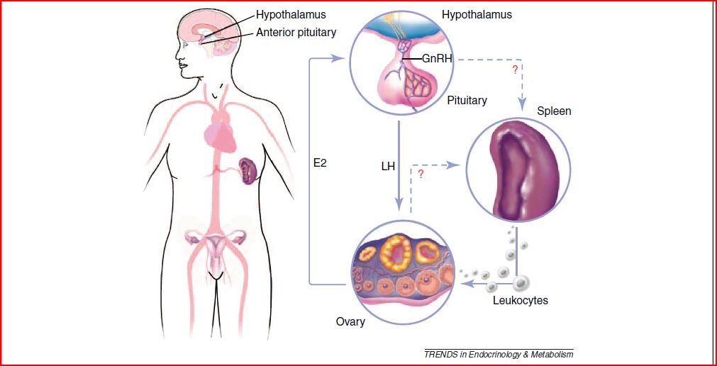
But how does this happen and where do these guys come from? The current hypothesis is that it happens via the same Hypothalamous-Pituitary-Ovarian axis that we should be familiar with but there's a twist to it and that's where the Spleen comes in. And isn't the spleen supposed to be some non functional purple organ that functions as a cell cementery in our bodies? Well, for females the answer now seems to be NO.

Recent studies, using the above mentioned flow cytometry have demostrated that after a heart attack, the spleen starts sending out armies of leukocytes to deal with the inflammation inside the heart. The same approach was used to asses the procedence of leukocytes arriving at the ovary during the  pre-ovulatory and ovulation period and they ended up seeing that as the ovarian vessels start filling up with these inflammatory cells, the spleen's population of leukocytes starts decreasing. Up to this point, it is thought that the LH hormone is the one responsible for going up to the hypothalamous and triggering the send me some white cells!!! signal moments prior to the start of ovulation and even if the above has been tested, it is in the triggering mechanism of this physiological inflammation where most questions remain.

There is a simple question that should arise from this: then does this mean that women who go trough splenectomy should be considered infertile? The problem is that along with splenectomy comes a process of radiotherapy and chimiotherapy wich could alone be harmful to the female reproductive abilities and it's difficult to measure this effect on humans. However, studies in animals who have gone trough splenectomy show that  the ovulatory becomes irregular and some of them actually stop ovulating all together. Another intriguin fact is that, considerably large leukocytes populations have been spotted in other female reproductive organs such as the uterus and flow cytometry shows that not all of the leukocytes coming out from the spleen end up in the ovary wich could also mean that at the same time they end up somewhere else in the reproductive tract.

Seems like we'll have to start thinking of ovulation as an important inflammatory event in no time and that's where the bridge between immunology and reproductive sciences will have to keep being crossed. This promises to be an interesting ride!
lunes, 29 de agosto de 2011

CPAP in newborns.

Before further ado, I'll go ahead and let you guys know that CPAP means Continuous Positive Airway Pressure.

First time I heard about it was regarding Sleep Apnea, that article expressed that CPAP was a valuable treatment for it, along with weight loss and other adjustments of course. Well, after a while of that and without hearing the word CPAP again, it was brought up today in our Pediatrics and Childcare class. 

It caught my attention cause our teacher mentioned that the childcare unit of our regional public hospital was going to received some of these devices. She was so excited while mentioning it and I just couldn't get it, I thought she was all "we're going to receive something new! Wee!" but after reading about the use and advantages of the CPAP in newborns that are either pre-term or just with breathing issues, I finally got her, most of all the potential advantages it has in developing countries like ours. This article explains how the use of CPAP for primary management of RDS (Respiratory Distress Syndrome) allows patients to save money  by not starting treatment with SRT (Surfactant Replacement Therapy) and instead using it in non-responders to CPAP 

Might sound unimportant to some people or just something already known and re-known for that matter, well yes, I'm not going to argue with that, it's not a new therapy so private clinics already have them. Usually money it's not an issue for those who attend to private clinics, they can pay for their services or they have a pretty good insurance but the reality of the poor part of society is completely different. Let me be breve and let you know about what public hospital means in "my language", it's a place/building the government has enabled with beds, some basic equipment (X-ray machine, EKG machine and others), doctors, nurses, residents, interns and med students.. everything else comes from your pocket, with that I mean "even the gauze" comes on your account. So, imagine that it must not be easy to have a newborn in NICU and be told what you need to do to keep your child alive but in the end you know you just don't have the means... 

360· and back to the essence of the subject, here are two pictures, one is a nasal-CPAP and the other is a helmet-CPAP, even if both of them do "the same" work, some authors have their preferences and the why's of it.   But I'm not planning to get into that,  here's and article that refers about it, there's more from where it came from =P, so I invite you to inform yourself a little about it. 

Good Night!


Test de APGAR / APGAR Score

Es un test utilizado en neonatología, que persigue la valoración clínica del neonato. Ideado e implementado en 1952 por Virginia Apgar, el test consta de la valoración de 5 parámetros en una escala de medición 0-2 cada uno, siendo la puntuación final de 0 a 10. Debe realizarse el test al minuto de nacer y luego a los 5 minutos, la primera vez permite valorar la torancia del proceso de nacimiento, mientras que la segunda permite la valoración de la adaptabilidad y capacidad de recuperación.

The Apgar scoring is used to evaluate the newborn in a general matter. Virginia Apgar introduced this test in 1952. In order to perform it one must evaluate 5 simple criteria on a scale from 0-2, then summing up the five values, the resulting Apgar score ranges from 0 to 10. One must perform this test before1 minute after birth and then again 5 minutes later, the first time we assess the tolerance of the process of birth and the second time we assess the ability to adapt and capacity of recovery. 

A - Appearance / Apariencia (Color of the skin / Color de la piel)
P - Pulse / Pulso (Cardiac Frequency / Frecuencia Cardiaca)
G - Grimace / Gesticulación (Reflexes / Reflejos)
A - Activity / Actividad  (Muscular Tone / Tono Muscular)
R - Respiration / Respiración (Quality not frequency)


Conociendo el significado de las puntuaciones obtenidas:
Una puntuación de 8-10 = Buenas condiciones.
Una puntuación de 4-6 = Se necesita vigilancia y valoración clínica.
Una puntuación <4 = Considerada grave, se requiere ventilación y medicamentos IV.
Una puntuación de 0 = Probable muerte fetal/neonatal. Junsto a un Ph igual o < 7.

Getting to know the meaning of the scoring obtained:
A scoring of 7-10 = Considered normal, good conditions. 
A scoring of 4-6 = Clinical watch and assessment needed.
A scoring below 4 = Considered an emergency, ventilation and IV medications required.
A scoring of 0 = Probable fetal/neonatal death. Along with a Ph lower or equal to 7.

Espero que disfrutaran la explicación y que la hayan comprendido, sino cualquier pregunta es bienvenida. Me parece que es buena idea tomarme el tiempo de escribir en ambos idiomas y así la información puede llegar a más personas =), ya el francés vendrá por ahí!.

I hope you guys enjoyed the explanation and most importantly understood it, if otherwise any question is welcome. I got to the conclusion that's a good idea to take some time and write both spanish and english, that way more people can get benefit from the information. Soon enough will come french =P.

Hasta la próxima / Until next time.

martes, 9 de agosto de 2011

Mini Mental Test de Folstein. Folstein Mini Mental State Exam(MMSE).

Es un test realizado principalmente en el área de neurología, con la finalidad de evaluar ciertos aspectos cognitivos del paciente, se basa en una puntuación de 30pts y puede realizarse en unos pocos minutos dependiendo de la experiencia con que cuente el evaluador.

El test consta de 11 pasos, una puntuación menor de 24 sugiere demencia; la misma tiene diferentes grados, entre 23-21 la demencia es leve, entre 20-11 nos encontramos frente a una demencia moderada y menor de 10 de una demencia severa. Aquí los puntos que debemos evaluar y cómo hacerlo.

1. Orientación en tiempo (5pts)
Preguntar al paciente:
- El día de la semana (1).
- Fecha  (número en el calendario) (1).
- Mes (1).
- Año (1).
- Estación del año (1).

2. Orientación en lugar (5pts)
Preguntar al paciente:
- Lugar de la entrevista (Planta o área del hospital) (1).
- Hospital (1).
- Ciudad (1).
- Provincia (1).
- País (1).

3. Registro de 3 palabras (3pts)
Se pide repetir 3 palabras, se otorga un punto por cada palabra que el paciente recuerde.

4. Atención y Cálculo (5pts)
Serie de 7. Pedir al paciente que reste 7 iniciando desde 100. Realizar 5 repeticiones, por cada correcta se otorga un punto. Ej. 100 - 7 = 93 - 7 = 86 - 7 = 79... Puede también pedirse al paciente que deletree la palabra MUNDO al revés. Ej. O - D - N - U - M.

5. Recall (3pts)
Se otorga un punto por cada palabra que el paciente recuerde de las 3 que se pidió memorizar en el paso 3.

6. Nominación (2pts)
Pedir al paciente que nombre dos objetos que serán mostrados, otorgar 1 punto por cada una que se responda correctamente.

7. Repetición (1pt)
Pedir al paciente que repita una de estas oraciones:
- Tres perros en un trigal.

8. Comprensión (3pts)
Indica al paciente que realice 3 acciones, por ejemplo: Tome este lápiz (1), colóquelo sobre la mesa (1), tómelo con la mano izquierda (1). O el clásico Tome este papel (1), dóblelo a la mitad (1), póngalo en el suelo (1).


9. Lectura (1pt)
Escribir una frase en un papel, pedir al paciente que la lea para él mismo y luego la realice. Ej. "Levante su mano derecha" o el clásico "Cierre los ojos"

10. Escritura (1pt)
Pedir al paciente que escriba una oración con sentido lógico.

11. Dibujo o destrezas visuoespaciales (1pt)
Realizar este dibujo y pedirle al paciente que lo copie al lado. Son dos pentágonos cruzados en las puntas.


Aquí les dejó unas páginas con unos buenos ejemplos y guías.
1. http://www.geroeducation.org/HyperText_Module/Delirium/html/mmse.htm
2. http://lifemanagement.com/nextsteps/Mini_Mental_Status_Exam.pdf
3. http://bit.ly/q4atrr

martes, 12 de abril de 2011

Clasificación TNM


Clasificación TNM explicada de forma sencilla y fácil de aprender. Extraída de la sección de Neumología del CTO.

domingo, 13 de marzo de 2011

Criterios Diagnósticos: Miocarditis

La Miocarditis es una condición de difícil diagnóstico, en la búsqueda de facilitar su rápido tratamiento y así mejorar la calidad de vida del paciente, siempre se buscan los mejores medios y criterios para lograr descubrirla. 

El diagnóstico patológico ha sido el único formalmente aprobado, por ende la biopsia es la prueba dx estándar. Los criterios Dallas constan siemplemente de dos hallazgos:
1. Presencia de células inflamatorias.
2. Necrosis del tejido celular. 

Otros criterios usados en clínica se muestran en esta tabla, extraída del libro de Cardiología de Braunwald 8ed. 


TABLE 66-2   -- Expanded Criteria for Diagnosis of Myocarditis
Suspicious for myocarditis = 2 positive categories
Compatible with myocarditis = 3 positive categories
High probability of being myocarditis = all 4 categories positive
(Any matching feature in category = positive for category)

   Category I: Clinical Symptoms
   Clinical heart failure
   Fever
   Viral prodrome
   Fatigue
   Dyspnea on exertion
   Chest pain
   Palpitations
   Presyncope or syncope

   Category II: Evidence of Cardiac Structural/Functional Perturbation in the Absence of Regional Coronary Ischemia
   Echocardiography evidence
   Regional wall motion abnormalities
   Cardiac dilation
   Regional cardiac hypertrophy
   Troponin release
   High sensitivity (>0.1 ng/ml)
   Positive indium-111 antimyosin scintigraphy
   and
   Normal coronary angiography or
   Absence of reversible ischemia by coronary distribution on perfusion scan

   Category III: Cardiac Magnetic Resonance Imaging
   Increased myocardial T2 signal on inversion recovery sequence
   Delayed contrast enhancement following gadolinium-DTPA infusion

   Category IV: Myocardial Biopsy—Pathological or Molecular Analysis
   Pathology findings compatible with Dallas criteria
   Presence of viral genome by polymerase chain reaction or in situ hybridization

Un dato útil:
Myocarditis as a diagnosis should be suspected when a young patient presents with unexplained symptoms of heart failure or chest pain, but the coronary arteries are found to be normal on angiography.

jueves, 10 de marzo de 2011

Endocarditis Infecciosa (EI) : Criterios de Duke

La EI, podría breve y superficialmente definirse como infección de la capa interna que recubre el corazón, llamada endocardio, que provoca una subsecuente inflamación y que afecta principalmente las válvulas cardíacas ( que están recubiertas por este endocardio ). La EI no se produce por la simple presencia de bacteremia, se necesitan una serie de requisitos para que ésta se desarrolle, como lo es el daño endotelial, patologías valvulares, alteraciones en la anatomía cardíaca, ect., pero siendo el principal factor la instauración previa de una Endocarditis Trombótica No Bacteriana ( NBTE ) sobre la cual se depositarán los agentes bacterianos o fúngicos presentes en la sangre durante dicha bacteremia.

Ésta NBTE es parte importante de la patogénesis de la EI, está formado principalmente de fibrina y se aloja en las válvulas cardíacas. Es también la explicación de la endocarditis en personas sin alteraciones de otro tipo ( como las mencionadas anteriormente ).

La EI es considera una condición algo incómoda de diagnosticar, sin embargo se han ido sumando métodos efectivos ante la sospecha. En la clínica los criterios de Duke son los más ultilizados, mientras que el ecocardiograma es el arma clave para confirmar la presencia de vegetaciones.

A continuación en el siguiente link podrán encontrar una tabla que contiene los criterios modificados de Duke, que permiten un diagnóstico más certero de EI.
https://blogger.googleusercontent.com/img/b/R29vZ2xl/AVvXsEgG_JyK1h3zPFIIR3HRynZJEv0vowuKqM2p67DkrJsirhvXktXlTzpfF0W1StnaRWannVakTvmyiqLoHM6igIqJoUSiuOlIo3Qw_Y9nL4NdACPF1BlHGRbrUV_Nb-2OU_B8JxoieGLj5A/s1600/Tabla+2.gif.

Puede establecerse un diagnóstico de endocarditis definitivo o un diagnóstico posible en base a los hallazgos encontrados en el paciente, basándonos claramente en estos criterios.

- Dx definitivo:
  1. Dos criterios mayores 
  2. Un criterio mayor y 3 menores
  3. Cinco criterios menores
- Dx posible:
  1. Un criterio mayor y un criterio menor
  2. Tres criterios menores